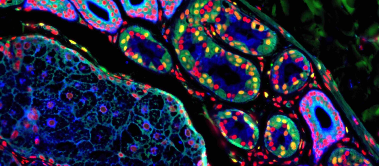
Skeletal muscle is a highly adaptable tissue with well-known sensitivities to environmental cues such as growth factors, cytokines, nutrients and mechanical loading. Moreover, adult skeletal muscle exhibits great plasticity and has the unique capacity to regenerate after injury, due to satellite cells, the muscle stem cells. Satellite cells, and their progeny, the myogenic precursor cells, must develop proper interactions with their direct environment to ensure muscle regeneration and replenishment of the stem cell pool. The aim of our research is to understand the role of the environment (non-myogenic cells and molecular structures) in the regulation of skeletal muscle regeneration. Myogenic cell environment includes inflammatory cells and particularly macrophages, vessel cells (endothelial and peri-endothelial cells) and interstitial cells. We aim at understanding the interactions between myogenic cells and their environment in various physiological and pathological situations.
Muscle stem cell neighboring (B. Chazaud)
Skeletal muscle regeneration is associated with the presence of macrophages. Two main types of macrophages are present during skeletal muscle regeneration. Soon after injury, inflammatory monocytes enter into the damaged muscle (1) and these inflammatory macrophages stimulate the proliferation of muscle stem cells (2). Upon efferocytosis, which is the engulfment of cell and tissue debris, they switch their phenotype into anti-inflammatory/repair macrophages (3), that sustain myogenic differentiation (4) fusion and myofiber growth (5), as well as angiogenesis and matrix remodeling, until the return to homeostasis (6). Macrophages can be considered as a major actor of the regenerative niche of myogenic cells that helps the sequential steps of skeletal muscle regeneration. An important aspect in macrophage functions is their dynamic during tissue repair, with a crucial moment when the inflammatory phase ends to start the repair process, that is the resolution of inflammation.
Neuromuscular electrical stimulation training to fight cachexia (J. Gondin)
Cachexia is a common consequence of many chronic diseases including sepsis and cancer. This loss of skeletal muscle mass ultimately results in increased morbidity and mortality. Moreover, treatments, sedation and/or prolonged unloading may further increase muscle deconditioning. On that basis, the development of countermeasures to prevent or attenuate the loss of skeletal muscle mass is of utmost importance for both patients. Our aim is to investigate whether and to what extent NeuroMuscular Electrical Stimulation (NMES) is a promising therapy for increasing force production and promoting muscle growth in preclinical models of cachexia. The program aims at establishing rigorous protocols to exploit the benefit of NMES on muscle function and at identifying the cellular and molecular events underlying those beneficial effects.
Hereditary ataxias are a heterogeneous set of severely disabling neurological disorders caused by degeneration of the cerebellum and/or the spinal cord. The prevalence of hereditary ataxias is estimated to 1/20,000 individuals in Europe, and yet there are no specific treatments for most of them.
Our research focuses on understanding the pathophysiology of ataxia, discovering disease biomarkers and developing therapeutic approaches. In addition, in close collaboration with the clinicians, we are developing new diagnostics tools for cerebellar ataxia and identify novel genes causing ataxia.
We are mainly interested in two different recessive ataxias, Friedreich ataxia (FA) and autosomal recessive cerebellar ataxia 2 (ARCA2), linked to two essential mitochondrial pathways: iron-sulfur cluster (ICS) biosynthesis and coenzyme Q10 (CoQ10) biosynthesis, respectively; FA belongs to the family of trinucleotide repeat disorders, which are caused by dynamic mutations that show instability (expansion/contraction) in the germline and in selective somatic cells.
Friedreich ataxia (FA), the most common recessive ataxia, is characterized by progressive gait and limb ataxia associated with hypertrophic cardiomyopathy and an increase incidence in diabetes. The major mutation is a GAA repeat expansion within the first intron of the FXN gene. In FA, the GAA expansion leads to heterochromatinization of the locus resulting in a drastic decrease of transcription of FXN. The disease results from loss of function of FXN gene product, frataxin, a highly conserved mitochondrial protein involved in the biogenesis of ISC, which are essential protein cofactors implicated in numerous cellular functions.
The autosomal recessive cerebellar ataxia 2 (ARCA2) is characterized by cerebellar ataxia and atrophy, and is associated with exercise intolerance. Most patients present a mild deficiency in CoQ10 in muscle biopsies. ARCA2 results from loss of function mutations in the ADCK3/COQ8A gene that encodes a mitochondrial protein with a regulatory role in CoQ10 biosynthesis.
Key words: ataxia, mitochondria, genetics, models (cell, mouse, zebrafish), therapeutic approaches
The central goal of our team is to understand how the excitability of neurons and muscles is regulated by basic molecular and cellular processes that control the biology of potassium-selective ion channels.
To identify novel genes and conserved cellular processes that regulate the biology of potassium channels in vivo, we take advantage of the powerful genetic tools available in the model nematode Caenorhabditis elegans. We use the full array of techniques available in C. elegans including genetics, live imaging, electrophysiology and state-of-the-art CRISPR/Cas9 genome engineering and next-generation DNA sequencing.
Our team studies the cellular and molecular basis sustaining neuronal integrity, and deciphers the mechanisms underlying neurodegeneration and/or developmental defects in disease.
Initially focused on a fatal neurodegenerative disease called GAN (giant axonal neuropathy), we identified the encoded Gigaxonin-E3 ligase and uncovered its pivotal roles in controlling cytoskeletal architecture (Intermediate Filaments), autophagy machinery (ATG16L1) and neuronal identity (Shh signaling through Ptch). Our scientific direction is to further tackle the biology of neuromuscular diseases. Specifically, we aim to unravel the regulations and functions of i) the cytoskeleton and ii) the autophagy pathway in normal neuronal physiology and disease states, and then translate this knowledge into therapies.
Our team is strongly engaged in the dissemination of science (fundamental & translational), and organizes/participates to numerous events for the public and patients.