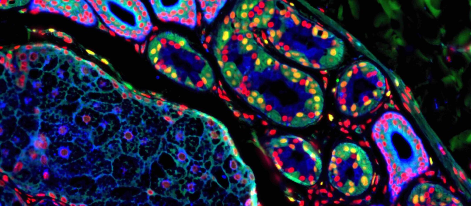
How do the immune system’s sentinels translate microbial and danger signals into immunological instructions?
Within the blood, mucosal tissues, and lymphoid organs, dendritic cells (DCs) fulfill a dual role as both sentinels and orchestrators of the immune system. Strategically positioned at pathogen entry points, they detect invading microorganisms, internalize them, and release biochemical mediators that alert the body’s defense networks and recruit antimicrobial effector cells to the site of infection. Following pathogen uptake and degradation, dendritic cells display antigenic fragments of the invader on their surface and subsequently migrate through lymphatic vessels to secondary lymphoid organs—including the spleen, lymph nodes, and mucosa-associated lymphoid tissues of the gastrointestinal and respiratory tracts. There, they present antigens to T and B lymphocytes, initiating adaptive immune responses. Once pathogen characteristics have been recognized, these lymphocytes—now equipped with highly specific and potent effector mechanisms—migrate to the infection site to ensure long-term clearance and immune memory.
The Laboratory of Dendritic Cell Biology (DeCiBeL) is dedicated to advancing the understanding of dendritic cell and phagocyte biology under both physiological and pathological conditions. Our mission combines fundamental research with the development of innovative tools to investigate the cellular and molecular mechanisms governing immune activation and regulation.
Our work focuses on the signaling pathways involved in the detection of microbe-associated molecular patterns (MAMPs). These pathways coordinate multiple cellular processes—such as inflammation, autophagy, intracellular trafficking, endoplasmic reticulum (ER) stress, and energy metabolism—to effectively activate and modulate immune responses. Among these mechanisms, autophagy and the unfolded protein response (UPR), a key component of ER stress regulation, are central to maintaining cellular homeostasis and linking inflammatory signaling to environmental stimuli implicated in autoimmune diseases and cancer.
We investigate how these interconnected pathways and translational control mechanisms fine-tune dendritic cell biochemistry and metabolism in response to diverse microbial and danger signals (MAMPs and DAMPs). The intricate crosstalk between these pathways necessitates the use of high-resolution quantitative methodologies, an area in which our laboratory actively contributes to technological development. These efforts aim to elucidate how dendritic cells acquire their specialized immunoregulatory functions, thereby informing novel therapeutic strategies in oncology and autoimmunity.
Dynamics of endomembrane systems in innate and adaptive immune detection and signaling
Recognition of microbial patterns profoundly influences the organization of intracellular membrane systems, particularly the endocytic network, thereby enabling dendritic cells to efficiently acquire immunostimulatory properties. Our research has demonstrated that the BAD-LAMP/LAMP5 protein is predominantly expressed in human plasmacytoid dendritic cells (pDCs) and in blastic plasmacytoid dendritic cell neoplasms (BPDCN). BAD-LAMP regulates the trafficking of TLR9 to signaling endosomes, thereby modulating pDC activation (Combes et al., Nature Communications, 2017). Furthermore, we have shown that BAD-LAMP is expressed in acute myeloid leukemia (AML) cells derived from patients, where it sustains cellular survival and tumor progression through activation of the NF-κB signaling pathway (Maldonado et al., 2022).
In parallel, we are elucidating the functions of the RUFY protein family in phagocytes (Char et al., 2020). RUFY proteins are characterized by a RUN domain, which interacts with small GTP-binding proteins, and a FYVE domain, responsible for recognizing PtdIns(3)P lipids within organelles. Beyond their established role in autophagy regulation within dendritic cells, our findings indicate that RUFY4 participates in the selective elimination of mitochondria in alveolar macrophages through effectors including PLEKHM1, Rab7, the HOPS complex, and various mitochondrial proteins (Valecka et al., 2022).
We also identified a dendritic cell–specific isoform of RUFY3, which contributes to lysosomal clustering in macrophages and DCs in response to bacterial lipopolysaccharide (LPS) (Char et al., 2023). Collectively, our results establish RUFY3 as a regulatory component of IFN-γ signaling, antigen presentation, and cell migration, with a demonstrable anti-inflammatory function in vivo. RUFY3 interacts with the small GTPase ARL8b, which governs dynein-mediated transport of late endosomes along microtubules and links this process to PtdIns(3)P synthesis mediated by the class III phosphatidylinositol 3-kinase Vps34. Complementary experiments have shown that pharmacological inhibition of Vps34 disrupts both RUFY3 distribution and TLR7/9 signaling, highlighting the interdependence of endomembrane dynamics and innate immune signaling pathways.